Exploring the Practical Applications of Blurring in Medical Imaging and Scientific Research
Blur: NFT | Blur: NFT login | Blur: NFT connect | WalletConnect | Traders | What Is Blur Crypto
Blur: NFT | Blur: NFT login | Blur: NFT connect | WalletConnect | Traders | What Is Blur Crypto
Blur, often associated with imperfections or errors, has found its place in various fields of science and technology. In medical imaging and scientific research, blur is no longer seen as a mere drawback, but rather as a valuable tool with numerous real-life applications.
One of the primary applications of blur in medical imaging is in the field of radiology. By intentionally introducing controlled blur, medical professionals are able to enhance certain details in radiographic images. This technique, known as motion-blur imaging, allows for improved visualization of organs and tissues, helping to identify and diagnose various conditions more accurately. As a result, medical practitioners are able to make informed decisions regarding patient treatment and care.
In scientific research, blur is also utilized for a wide range of applications. For example, in microscopy studies, blur can be intentionally introduced using specific lenses or techniques to overcome the limitations of traditional imaging methods. This blurring effect allows for the capture of high-resolution images of microscopic structures, enabling scientists to study cellular processes and interactions in greater detail. The insights gained from such studies contribute to advancements in various fields, including biology, chemistry, and medicine.
Moreover, the utilization of blur in scientific research extends beyond microscopy. In fields such as astronomy and astrophysics, astronomers intentionally induce blur to study distant celestial objects. By analyzing the effects of atmospheric turbulence, researchers are able to understand and correct for the blurring caused by Earth's atmosphere. This correction allows for the production of clearer and more detailed images, providing astronomers with invaluable insights into the universe and its mysteries.
In conclusion, the intentional utilization of blur in medical imaging and scientific research has revolutionized these fields by introducing new capabilities and insights. From motion-blur imaging in radiology to the deliberate introduction of blur in microscopy and astronomy, scientists and medical professionals are harnessing blur's potential to enhance and improve their studies. As technology continues to advance, the applications of blur are expected to expand further, leading to even greater breakthroughs and advancements in various scientific disciplines.
The Application of Blur in Medical Imaging and Scientific Research
The utilization of blur has proven to be a significant tool in medical imaging and scientific research. This technique has been employed to enhance the accuracy and quality of various imaging modalities, such as X-rays, computed tomography (CT), magnetic resonance imaging (MRI), and ultrasound. The application of blur in medical imaging allows researchers and doctors to obtain clearer and more detailed images, which can aid in the diagnosis and treatment of various medical conditions.
One of the main benefits of utilizing blur in medical imaging is the reduction of image noise and artifacts. Image noise can occur due to various factors, such as sensor noise, patient movement, or external interference. By applying blur to the image, these noise and artifacts can be minimized, resulting in a clearer and more accurate representation of the underlying anatomy or pathological features.
Furthermore, the application of blur can help in the visualization and analysis of complex anatomical structures. For example, in CT or MRI scans, blur can be used to enhance the edges and boundaries of different organs or tissues, making it easier for radiologists and researchers to identify and measure specific structures of interest. This can be particularly useful in surgical planning and monitoring, as well as in the study of anatomical variations and abnormalities.
In addition to medical imaging, the application of blur has also found its way into scientific research in various fields. For instance, in microscopy, blur can be used to improve image resolution and depth of field, allowing researchers to observe and analyze microscopic structures with greater clarity and precision. This can have implications in fields such as cell biology, genetics, and materials science.
Moreover, the recent advancements in artificial intelligence and machine learning have further expanded the possibilities of utilizing blur in medical imaging and scientific research. Algorithms and models can be trained to automatically deblur images, improving the overall quality and diagnostic value of medical and scientific images. This has the potential to save time and resources, as well as improve the accuracy and efficiency of diagnosis and analysis.
In conclusion, the application of blur has proven to be an invaluable tool in medical imaging and scientific research. With its ability to reduce image noise, enhance anatomical structures, and improve image resolution, blur has the potential to revolutionize the way we acquire, analyze, and interpret medical and scientific images. To learn more about the applications and advancements in blur technology, visit Blur: NFT.
Understanding the Concept of Blur

Blur is a term widely used in the field of medical imaging and scientific research to describe the loss of sharpness or resolution in an image. It occurs when the edges of objects in an image become less defined and appear to blend together, resulting in a decrease in visual acuity.
In medical imaging, blur can be caused by a variety of factors. One common cause is the movement of the patient during the image acquisition process. When a patient moves, it can introduce blur into the image, making it more difficult for healthcare professionals to accurately interpret the results. This is particularly problematic in imaging modalities such as X-rays, where high precision and clarity are crucial for accurate diagnosis.
Another cause of blur in medical imaging is the limited resolution of the imaging equipment itself. Each imaging modality has its own inherent resolution limit, beyond which the details in the image can become blurred. For example, in magnetic resonance imaging (MRI), the voxel size determines the resolution of the image, and if the voxel size is too large, the image may appear blurry.
Blur in Scientific Research
Blur also plays a significant role in scientific research. In microscopy, for example, blur can occur due to limitations in the optics of the microscope or the properties of the specimen being observed. This can hinder researchers' ability to accurately study and analyze microscopic structures, such as cells or tissues.
Furthermore, blur can be intentionally introduced in scientific research to achieve specific effects. In some cases, blurring or distorting an image can provide insights into certain phenomena or facilitate data analysis. For example, in computer vision research, blur can be used to simulate the effects of motion blur, depth-of-field blur, or other optical phenomena, in order to develop algorithms or evaluate the performance of image processing techniques.
The Impact of Blur on Image Analysis
Understanding the concept of blur is crucial in the field of medical imaging and scientific research, as it directly affects the accuracy and reliability of image analysis. By recognizing and addressing sources of blur, researchers and healthcare professionals can improve the quality of images, enabling better diagnosis, analysis, and understanding of the underlying phenomena.
The Role of Blur in Medical Imaging

In the field of medical imaging, blur plays a crucial role in capturing accurate and detailed images of the human body. It is used intentionally to enhance certain aspects of the images, providing valuable information to medical professionals for diagnosis and treatment purposes.
One major application of blur in medical imaging is in the technique known as computed tomography (CT scan). CT scans involve taking multiple X-ray images from different angles and using computer algorithms to reconstruct a three-dimensional image of the patient's body. By intentionally introducing a controlled amount of blur in the images, the resolution and clarity of the reconstructed image can be improved. This is particularly useful in capturing fine details of structures such as blood vessels or small tumors, which might be difficult to visualize without the use of blur.
Blur is also utilized in medical imaging to optimize the visualization of certain anatomical structures. For example, when imaging the heart using echocardiography or cardiac MRI, intentionally inducing a slight blur can help reduce artifacts caused by motion, resulting in clearer images of the heart's structure and function. This allows medical professionals to more accurately assess the health of the heart and diagnose any underlying conditions.
Furthermore, in the field of ophthalmology, blur is commonly used to evaluate visual acuity and diagnose refractive errors. By presenting patients with blurred images and assessing their ability to perceive details, eye care professionals can determine the level of clarity and focus a patient's eyes possess. This information is crucial when prescribing corrective lenses or performing vision correction surgeries.
Overall, the controlled utilization of blur in medical imaging plays a vital role in enhancing image quality, improving diagnostic accuracy, and aiding in scientific research. Its application in various imaging techniques and disciplines contributes to advancing medical knowledge and improving patient care.
Enhancing Image Quality Through Blur
Blur, a commonly encountered phenomenon in medical imaging and scientific research, can actually be utilized to enhance image quality rather than being considered a hindrance. By deliberately introducing controlled levels of blur, researchers and medical professionals can obtain more precise and accurate results in various applications.
1. Noise Reduction:
The introduction of controlled blur can effectively reduce noise in medical images and scientific research. Noise, often caused by factors such as sensor limitations or environmental conditions, can affect the accuracy of measurements and analysis. By intentionally blurring the image, the noise can be smoothed out and minimized, resulting in cleaner and more reliable data.
2. Edge Enhancement:
Blur can be strategically applied to enhance the visibility of edges and boundaries in an image. In medical imaging, this can be particularly useful for highlighting anatomical structures or abnormalities. By judiciously blurring the surrounding areas, the edges become more prominent and easier to distinguish, enabling a more comprehensive and accurate diagnosis.
3. Detail Enhancement:
Contrary to intuitive perception, blur can also contribute to detail enhancement. By selectively blurring certain areas of the image, the finer details in other areas can be accentuated. This technique is commonly employed in scientific research to emphasize specific regions of interest or highlight subtle features that would otherwise be imperceptible.
4. Resolution Enhancement:
In situations where the image resolution is limited or restricted, introducing controlled blur can actually help enhance the perceived resolution. By averaging out the pixel values through controlled blurring, image details that were previously indistinguishable due to low resolution can become more discernible and provide valuable insights for analysis and interpretation.
In conclusion, blur should not be merely considered an undesirable artifact in medical imaging and scientific research. Instead, it can be harnessed as a valuable tool to enhance image quality and improve the accuracy and reliability of data analysis. By understanding the applications and employing deliberate strategies, researchers and medical professionals can leverage the benefits of blur to achieve more robust and informative results.
Blurring Techniques in Medical Imaging
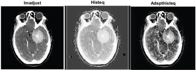
Blurring techniques are widely used in medical imaging to enhance the quality of images and improve diagnostic accuracy. By introducing controlled blurring or smoothing effects, medical professionals can reduce noise, enhance contrast, and highlight specific features of interest.
One common blurring technique used in medical imaging is Gaussian blurring. This technique involves applying a convolution operator to the image, which averages the intensity values of neighboring pixels and creates a smoother appearance. Gaussian blurring is particularly effective in reducing high-frequency noise and improving image clarity.
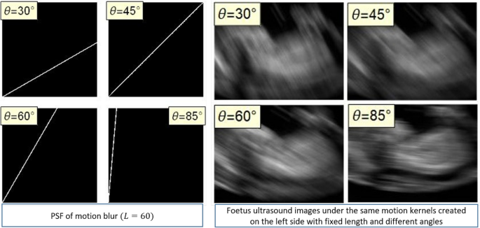
Another blurring technique used in medical imaging is motion blurring. This technique simulates the effect of motion by deliberately introducing a blur along a specific direction. Motion blurring can be used to improve the visibility of moving structures, such as blood vessels or organs during dynamic imaging procedures.
Applications of Blurring Techniques in Medical Imaging
Blurring techniques in medical imaging have several practical applications. One application is in the field of radiology, where blurring can be used to improve the visibility of internal structures, enhance tissue contrast, and aid in the detection of abnormalities.
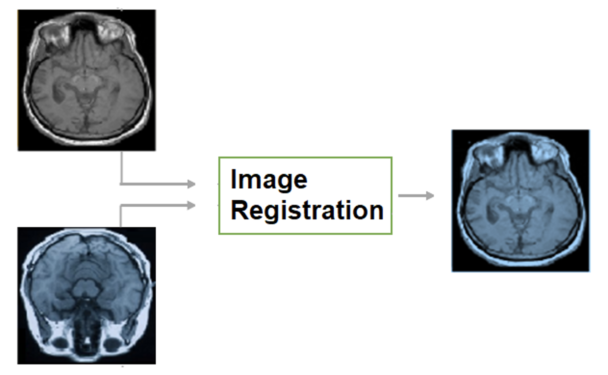
Another application is in image fusion, where multiple medical images from different modalities are combined to provide a comprehensive view of a patient's condition. Blurring techniques can be used to ensure seamless integration of images and alignment of anatomical structures.
Blurring techniques are also valuable in the field of image registration, where multiple images taken at different times or angles are aligned to create a coherent image. By incorporating blurring, the registration process can be improved by minimizing spatial mismatch and improving overall image quality.
1. Noise reduction
1. Loss of fine anatomical details
2. Contrast enhancement
2. Potential loss of diagnostic information
3. Highlighting specific features
3. Subjectivity in selecting blurring parameters
Overall, blurring techniques play a critical role in medical imaging by improving image quality, aiding in diagnostics, and facilitating image analysis and interpretation.
Blur's Contribution to Scanning Electron Microscopy

Scanning Electron Microscopy (SEM) is a powerful imaging technique widely used in scientific research and various fields of study, including materials science, biology, and nanotechnology. It allows scientists and researchers to visualize samples at high magnification and obtain detailed information about their surface structure and composition.
One of the challenges in SEM imaging is to ensure clear and crisp images with high levels of detail. This is where blur, specifically controlled blur, can contribute significantly to the image acquisition process. By intentionally introducing blur in SEM imaging, scientists can enhance certain features or reduce noise, thereby improving the overall quality and interpretability of the images.
Blur in SEM can be achieved through various methods, such as adjusting the focal plane, manipulating the electron beam, or employing special techniques like tilt photography. These techniques allow researchers to selectively blur specific areas or control the depth of field, resulting in images that provide a better understanding of the sample's structure and composition.
Blur also plays a crucial role in reducing artifacts and enhancing contrast in SEM images. By strategically applying blur, scientists can minimize the influence of unwanted reflections, shadows, or charging effects, which often occur during SEM imaging. This helps to produce more accurate representations of the sample and ensures that observed features are not distorted or obscured by artifacts.
The implementation of blur in SEM imaging is an area of active research and development. With advancements in image processing techniques, researchers can now introduce controlled blur digitally, allowing for more precise and flexible manipulation of the image data. This opens up new avenues for image enhancement and analysis, enabling scientists to extract valuable information that might have been overlooked in traditional SEM imaging.
Blur's contribution to scanning electron microscopy has proven to be invaluable in improving the quality and usability of SEM images. Its ability to enhance features, reduce artifacts, and provide better clarity has made it an essential tool for researchers in various scientific disciplines. As the field continues to evolve, the application of blur in SEM will undoubtedly play a central role in furthering our understanding of the microscopic world.
Applications of Blur in Electron Microscopy

Electron microscopy is a powerful technique used in scientific research to study the structure of materials at the atomic or molecular level. One interesting application of blur in electron microscopy is its use in enhancing resolution and reducing noise.
1. Resolution enhancement

Blur can be intentionally introduced in electron microscopy to improve the resolution of the images obtained. This technique, known as "defocus imaging", involves deliberately placing the image plane slightly out of focus. By doing so, the details of the specimen can be better visualized, as blur spreads out the diffraction patterns created by the electrons passing through the material.
Defocus imaging has been particularly useful in studying complex biological structures, such as proteins or viruses, where high resolution is crucial for understanding their functions and interactions. By carefully adjusting the degree of defocus, researchers can achieve a balance between increased resolution and acceptable image contrast.
2. Noise reduction
In electron microscopy, noise can arise from various sources, including the inherent electronic noise of the detector or the statistical nature of the electron interactions with the specimen. Blur can be employed as a tool to mitigate some of this noise, providing clearer and more informative images.
One common technique used to reduce noise is called "image averaging". By averaging multiple slightly blurred images of the same specimen, the random noise is effectively averaged out, while the true signal remains. This approach has been widely used in electron microscopy to improve the signal-to-noise ratio and reveal subtle details that would otherwise be obscured by noise.
Additionally, blur can also be utilized as a regularization method in image reconstruction, where the goal is to obtain a high-quality image from incomplete or noisy data. By intentionally blurring the image during the reconstruction process, artifacts and noise can be suppressed, resulting in a more accurate representation of the underlying structure.
In conclusion, the application of blur in electron microscopy has proven to be a valuable tool for enhancing resolution and reducing noise in scientific research. Whether it is used to improve the visualization of biological structures or to improve image quality in reconstruction algorithms, blur has demonstrated its usefulness in pushing the boundaries of what can be observed and understood at the nanoscale.
Using Blur to Study Particle Dynamics
The utilization of blur in medical imaging and scientific research extends beyond its applications in diagnosing and analyzing diseases. It can also be a powerful tool for studying particle dynamics in various fields of research.
When particles move rapidly or unpredictably, it can be challenging to capture their exact positions and movements with clarity. This is where blur comes into play. By intentionally introducing blur into images of particles, researchers can gain valuable insights into their dynamics, velocities, and interactions.
This technique is particularly useful in studying fluid dynamics, where particles suspended in a liquid or gas exhibit complex and often chaotic behaviors. By intentionally blurring the images of these particles, researchers can analyze the patterns and trajectories of their movements, allowing them to understand the underlying physics and phenomena at play.
One example of using blur to study particle dynamics is in the field of colloidal science. Colloidal suspensions, consisting of particles in a liquid medium, play a crucial role in various industries, including pharmaceuticals, materials science, and food technology. By intentionally blurring the images of colloid particles under different conditions, researchers can study their aggregation, sedimentation, and diffusion properties, which ultimately helps in developing new materials and optimizing industrial processes.
In addition to fluid dynamics and colloidal science, blur is also employed in other areas of research, such as studying the behavior of microscopic organisms, understanding the movement of pollutants in natural environments, and analyzing the dynamics of nanoparticles for drug delivery systems.
Overall, the utilization of blur in scientific research allows researchers to go beyond traditional imaging techniques and delve deeper into the complexities of particle dynamics. By intentionally introducing blur, researchers can gain valuable insights and uncover hidden patterns, ultimately leading to advancements in various scientific and industrial fields.
Blur's Impact on Biological Imaging
In the field of biological imaging, blur plays a significant role in the acquisition and analysis of various types of images. This phenomenon, caused by factors such as motion, defocus, and aberrations, affects the quality and accuracy of images used in medical research and diagnosis.
Understanding Blur in Biological Imaging
Blur in biological imaging refers to the loss of sharpness and clarity in an image. It occurs when the imaging system fails to accurately focus on the target object or when there is motion during the image capture process. In both cases, the result is a blurred image that can hinder further analysis and interpretation.
Defocus blur occurs when the imaging system does not achieve optimal focus on the specimen. This can happen due to various reasons, including improper setup of the imaging equipment, limitations of the optical system, or material properties of the specimen itself. Defocus blur can make it difficult to discern fine details and accurately measure features of interest.
Motion blur, on the other hand, is caused by movement of either the imaging system or the specimen being imaged. In biological imaging, motion blur can be particularly problematic as biological samples are often dynamic and can move during the image acquisition process. This can result in images that are distorted and lack proper representation of the sample's true structure or function.
The Impact on Biological Research and Diagnosis
The presence of blur in biological images can have significant implications for both research and clinical diagnosis. Researchers heavily rely on clear and sharp images to analyze cellular structures, observe cellular processes, and study the effects of various treatments or interventions. Any blur in the images can lead to misinterpretations and inaccurate conclusions, potentially impacting the validity and reproducibility of the research findings.
In a clinical setting, accurate and detailed imaging is crucial for making diagnoses and determining appropriate treatment plans. Blur in medical images can obscure important anatomical features or artifacts, leading to misdiagnosis or delayed treatment. Additionally, it can affect the precise measurement of lesions, tumor boundaries, and other quantitative parameters that help in evaluating disease progression or treatment response.
The Need for Blur Reduction Techniques
To mitigate the impact of blur in biological imaging, various techniques and technologies have been developed. These include image deblurring algorithms, adaptive optics, motion compensation methods, and improvements in imaging hardware. By minimizing or eliminating blur, these advancements help improve the resolution, accuracy, and overall quality of biological images.
In conclusion, blur in biological imaging poses challenges for both researchers and clinicians. Understanding the causes and consequences of blur is crucial for developing effective strategies to reduce its impact and enhance the reliability and efficacy of biological imaging in a wide range of applications.
Utilizing Blur to Analyze Tissue Structures
In the field of medical imaging and scientific research, the utilization of blur has proven to be a valuable tool in analyzing tissue structures. By intentionally introducing controlled blur into imaging techniques, researchers are able to enhance certain features of tissues and gain a better understanding of their structures.
One of the main applications of utilizing blur in tissue analysis is in the field of pathology. Pathologists use blur as a technique called "optical clearing" to improve the transparency of tissues, making it easier to visualize and study their internal structures. This technique involves immersing the tissue in a clearing agent that has a refractive index closely matching that of the tissue's components. The resulting blur helps to reduce light scattering within the tissue, allowing for a clearer view of cellular and connective structures.
Another application of blur in tissue analysis is in the field of microscopy. Microscopic techniques often rely on precise focusing to capture detailed images of tissues. However, certain tissue structures may be difficult to distinguish or visualize clearly. By intentionally introducing controlled blur, researchers can enhance the visibility of specific features, such as cell boundaries or protein distribution, making it easier to analyze and study the tissue at a microscopic level.
In addition to pathology and microscopy, blur is also utilized in techniques such as optical coherence tomography (OCT). OCT is a non-invasive imaging technique that uses light waves to capture detailed cross-sectional images of tissues. By intentionally introducing blur, researchers can enhance the contrast between different tissue layers, allowing for a more accurate analysis of tissue structures. This can be particularly useful in diagnosing various medical conditions and monitoring the effectiveness of treatment.
Overall, the utilization of blur in tissue analysis has revolutionized the field of medical imaging and scientific research. By intentionally introducing controlled blur, researchers are able to enhance the visualization and analysis of tissue structures, leading to advancements in diagnosis, treatment, and understanding of various diseases and conditions.
Blur's Importance in Optical Coherence Tomography
Optical Coherence Tomography (OCT) is a non-invasive imaging technique widely used in medical and scientific research. It provides high-resolution cross-sectional images of biological tissues, allowing for the visualization of microscopic structures and the detection of abnormalities.
The Role of Blur in OCT
Blur, or the intentional defocusing of OCT images, plays a crucial role in enhancing the quality and usefulness of OCT scans. By deliberately introducing controlled blur into the system, the sensitivity and resolution of the images can be improved, enabling a more accurate analysis of the tissue structures.
Blur in OCT is achieved by manipulating the coherence length of the light source used in the imaging system. The coherence length determines the axial resolution of the OCT images, which refers to the ability to distinguish between two closely spaced structures along the depth axis. By carefully adjusting the blur level, the resolution can be optimized to achieve the desired balance between image sharpness and sensitivity.
Advantages of Controlled Blur in OCT
Controlled blur in OCT offers several advantages in medical imaging and scientific research:
Improved Depth Resolution: By introducing controlled blur, the depth resolution of OCT images can be enhanced, allowing for better visualization and measurement of structures within tissues. This is especially crucial in ophthalmology, where precise imaging of the retinal layers is necessary for diagnosing and monitoring various eye diseases.
Enhanced Contrast: Controlled blur can also improve contrast in OCT images, making it easier to differentiate between different tissue types or identify subtle changes within a tissue. This is particularly important in cancer research, where early detection of malignant cells relies on the ability to detect minute differences in tissue morphology.
Reduced Artifacts: In some cases, OCT images can suffer from artifacts, such as speckle noise or shadowing effects. Controlled blur can help mitigate these artifacts by smoothing out irregularities in the image and reducing noise, resulting in cleaner and more accurate representations of the tissue structure.
Overall, the utilization of blur in OCT plays a crucial role in improving image quality, resolution, and sensitivity, enabling researchers and medical professionals to obtain more detailed and accurate information about biological tissues. This, in turn, aids in the diagnosis, treatment, and understanding of various diseases and conditions.
Blur's Role in Microscopic Imaging Techniques

In the field of scientific research, microscopic imaging techniques play a crucial role in examining and understanding various biological structures and processes. One important aspect of these techniques is the utilization of blur, which can provide valuable information about the microscopic specimens being examined.
Blur, or image blurring, refers to the effect of making an image appear less sharp and clear. While blur is generally seen as undesirable in traditional photography, it is intentionally introduced in microscopic imaging techniques to enhance specific features or gather additional information.
One such application of blur is in the technique of confocal microscopy. In confocal microscopy, a pinhole or a slit is placed in front of the detector, which allows only a thin section of the specimen to be in focus while the rest of the image is intentionally blurred. This selective blurring helps eliminate the unwanted light originating from out-of-focus planes, resulting in a sharper and clearer image of the desired focal plane.
Another application of blur in microscopic imaging is in the technique of structured illumination microscopy (SIM). SIM utilizes structured light patterns to capture multiple images of a specimen with slightly shifted illumination patterns. By intentionally blurring each of these images, it becomes possible to extract fine details beyond the diffraction limit, thus achieving higher resolution imaging.
Furthermore, blur is also used in techniques such as deconvolution microscopy, where a blurred version of the acquired image is computationally processed to reconstruct a clearer and more detailed representation of the specimen. This deblurring process helps overcome the limitations imposed by factors such as diffraction, noise, and optical aberrations.
In summary, blur plays a significant role in various microscopic imaging techniques. It can be intentionally introduced to enhance certain features, eliminate unwanted light, surpass diffraction limits, and improve overall image clarity. By understanding and utilizing blur in these techniques, scientists and researchers can further advance their understanding of biological structures and processes at the microscopic level.
Applications of Blur in Confocal Microscopy
Confocal microscopy is a powerful imaging technique used in various fields of scientific research and medical diagnostics. It involves capturing images layer by layer, allowing for a deeper understanding of cellular structures and functions. Blur, although often considered undesirable in traditional imaging, plays a crucial role in confocal microscopy. Here are some of the applications of blur in this technique:
1. Depth Determination
Confocal microscopy uses a pinhole to block out-of-focus light and only allows light from the focal plane to reach the detector. This creates a blur in the acquired image, where each point appears as a small disc due to the out-of-focus blur. By studying the size of these discs, researchers can determine the depth of specific structures within a specimen. This depth information is essential for understanding the three-dimensional organization of biological samples.
2. Resolution Enhancement
Confocal microscopy can achieve higher resolution than conventional microscopy techniques by taking advantage of blur. When an object is beyond the resolution limit of a microscope, it appears as a blur due to diffraction. By limiting the detection to the focal plane, confocal microscopy reduces the blur caused by diffraction, resulting in enhanced resolution. This enables researchers to observe fine details and structures that would otherwise remain invisible.
Furthermore, using blur to enhance resolution allows for the removal of scattered light interference, improving image contrast and quality. By selectively collecting in-focus light, confocal microscopy minimizes the noise caused by scattered light signals, leading to sharper and more accurate images.
In conclusion, blur, once considered an obstacle in imaging, has proven to be a valuable asset in confocal microscopy. Its applications in depth determination and resolution enhancement have revolutionized our understanding of cellular structures and functions. As advances in technology continue to refine confocal microscopy, the role of blur will undoubtedly play an even more significant role in future scientific discoveries and medical breakthroughs.
Exploring the Benefits of Blur in Magnetic Resonance Imaging
Medical imaging techniques play a crucial role in diagnosing and monitoring various conditions. One such technique is Magnetic Resonance Imaging (MRI), which uses powerful magnets and radio waves to generate detailed images of the body's internal structures. In recent years, researchers have started exploring the benefits of intentionally introducing blur into MRI images to enhance their diagnostic value.
Improved Visualization of Fine Structures
By intentionally introducing controlled blur into MRI images, researchers have found that they can improve the visualization of fine structures within the body. This is especially useful in imaging complex anatomical regions where different tissues may closely interact. The controlled blur can help differentiate between these structures and provide a clearer depiction of the area of interest.
Enhanced Detection of Abnormalities
Blur can also be utilized to enhance the detection of abnormalities within MRI images. In certain cases, subtle abnormalities may be difficult to discern in crisp, high-resolution images. By introducing controlled blur, these abnormalities can be accentuated, making them more conspicuous and easier to detect. This can lead to earlier identification and treatment of various conditions.
Reduced Noise and Artifacts
Noise and artifacts are common challenges in medical imaging, including MRI. These can hinder the accuracy and reliability of the generated images, making interpretation more challenging. Intentionally introducing blur can help reduce the effects of noise and artifacts, resulting in cleaner and more reliable images for analysis. This can enhance the overall quality of MRI scans and improve the confidence in their diagnostic value.
Improved Patient Comfort
Another benefit of blur in MRI is improved patient comfort during the scanning process. MRI scans often require patients to remain still for extended periods. However, even slight movements can introduce motion artifacts into the images, compromising their quality. By intentionally introducing blur, the sensitivity to patient motion can be reduced, allowing for more flexibility in patient positioning and potentially improving patient comfort.
Conclusion
Exploring the benefits of blur in Magnetic Resonance Imaging has opened up new possibilities for improving the diagnostic value and overall quality of MRI scans. Through controlled blur, researchers have found improved visualization of fine structures, enhanced detection of abnormalities, reduced noise and artifacts, and improved patient comfort. These advancements have the potential to revolutionize the field of medical imaging and provide better outcomes for patients.
Utilizing Blur for Image Deblurring Techniques
Blur is a commonly encountered problem in imaging technology, where the details and sharpness of an image are lost due to various factors such as motion, camera limitations, or poor focus. Fortunately, advancements in image processing techniques have led to the development of powerful deblurring algorithms, which aim to restore the clarity and sharpness of blurred images.
Understanding image blur
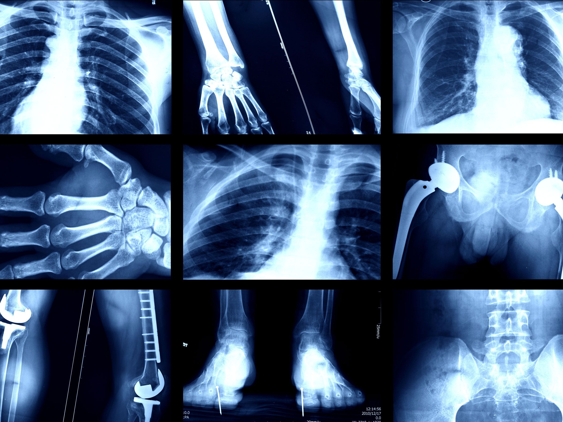
Image blur can be caused by different factors:
Motion blur: This occurs when there is relative motion between the camera and the subject during image capture. It often leads to a smearing effect.
Defocus blur: This type of blur is caused by incorrect focusing of the camera lens, resulting in a loss of sharpness and clarity.
Optical aberrations: These are lens imperfections that can introduce distortions and blurring effects to the final image.
Deblurring techniques
Several deblurring techniques have been developed to tackle the problem of image blur:
Blind deconvolution: This approach aims to recover the original image by estimating the blur kernel and the latent image simultaneously, without any prior knowledge of the blur characteristics.
Non-blind deconvolution: Unlike blind deconvolution, this technique requires prior knowledge of the blur kernel. It estimates the latent image by deconvolving the blurred image with the known kernel.
Image fusion: This method combines multiple blurred images of the same scene to create a high-quality, sharp image. It takes advantage of the fact that different images may have different blur patterns and combines their information to reduce blur.
Deep learning-based deblurring: This recent approach utilizes neural networks to learn the mapping between blurry and sharp images. It has shown promising results in restoring the details and sharpness of blurred images.
These deblurring techniques have found applications in a wide range of fields, including medical imaging, astronomical observations, microscopy, and forensics. They have significantly improved the quality of images and allowed researchers to extract valuable information that was previously obscured by blur.
In conclusion, the utilization of blur in image deblurring techniques has revolutionized the field of imaging technology. By understanding the causes of image blur and employing advanced deblurring algorithms, researchers and medical professionals can enhance the clarity and sharpness of images, enabling more accurate analysis and diagnosis.
Blur's Contribution to Image Restoration
Blur, typically seen as an undesirable effect in traditional photography, has proven to be an important tool in the field of image restoration, particularly in medical imaging and scientific research. By deliberately inducing controlled blur in images, researchers have been able to extract valuable information and enhance the quality of the original image using sophisticated algorithms and techniques.
One area where blur has made significant contributions to image restoration is in the enhancement of microscopic images. Microscopy techniques often suffer from inherent limitations in resolution and focus, leading to blurred or distorted images. However, by applying specific blur patterns and algorithms, it is possible to improve the clarity and sharpness of these images, revealing important details that were previously hidden or obscured.
Furthermore, blur has been extensively used in medical imaging, such as X-rays and CT scans, to reduce noise and enhance image quality. In these imaging modalities, noise can often degrade the visibility of critical structures or introduce artifacts that may lead to misdiagnosis. By strategically applying blur, noise can be minimized while preserving essential anatomical information, resulting in clearer and more accurate images for medical professionals to analyze and interpret.
In the field of scientific research, blur has played a crucial role in various applications, including astrophysics and particle physics. For instance, in astrophysics, atmospheric turbulence can cause blur in astronomical images, making it challenging to observe and study distant celestial objects. By implementing image deconvolution algorithms that take into account the blur caused by the Earth's atmosphere, scientists can reconstruct high-resolution images, enabling a deeper understanding of celestial phenomena.
Overall, blur's contribution to image restoration cannot be understated. It has revolutionized the field of medical imaging and scientific research, allowing for the extraction of valuable information from inherently flawed or degraded images. By embracing blur as a tool rather than disregarding it as a flaw, researchers and professionals can unlock new possibilities and insights in their respective fields.
To explore the cutting-edge advancements in image restoration and the utilization of blur, visit BLUR.IO アカウントへのログイン方法.
Future Potential of Blur in Medical Imaging and Scientific Research
The utilization of blur techniques in medical imaging and scientific research has already proven to be highly beneficial in various applications. However, its future potential is even more promising, with new advancements and possibilities on the horizon.
Improved Image Visualization
One area where blur can have a significant impact in the future is in improving image visualization in medical imaging. By selectively applying blur to specific areas of an image, researchers can enhance the visibility and clarity of certain structures or features. This can lead to more accurate diagnoses, better treatment planning, and improved patient outcomes.
For example, in neuroimaging, the use of blur can help highlight specific brain regions or abnormalities, making it easier for doctors to identify and analyze them. Similarly, in radiology, the application of blur can enhance the visibility of certain anatomical structures, aiding in the detection of diseases or abnormalities.
Simulating Real-life Conditions
Another potential application of blur in the future is in simulating real-life conditions for research purposes. By introducing controlled blurring effects into medical imaging or scientific experiments, researchers can create more realistic and representative environments.
For instance, in ophthalmology research, blur can be utilized to simulate various visual impairments, such as cataracts or glaucoma, allowing scientists to study their effects on visual perception and develop better treatment strategies. In the field of virtual reality, blur can be used to simulate depth-of-field effects, improving the immersion and realism of virtual environments.
Enhanced image visualization
Improved diagnoses and treatment planning
Better understanding of real-life conditions
Simulating visual impairments and environmental effects
How is blur used in medical imaging?
Blur is commonly used in medical imaging to remove fine details and focus on the overall structure. This can be helpful when conducting diagnoses or analyzing larger patterns within the images.
What are some real-life applications of using blur in medical imaging?
One real-life application of using blur in medical imaging is for the detection of tumors. By applying blur to an image, the surrounding healthy tissue can be deemphasized, making the tumor stand out more clearly. Another application is in cardiac imaging, where the use of blur can smooth out motion artifacts and improve the visibility of certain structures.
Are there any disadvantages to using blur in medical imaging?
While using blur in medical imaging can be advantageous in some cases, it can also result in the loss of important details. If too much blur is applied, it may become difficult to accurately interpret the images and make precise diagnoses. Additionally, the process of applying blur can be time-consuming and may require specialized software or techniques.
How is blur utilized in scientific research?
In scientific research, blur can be used to enhance the visibility of certain features or to simulate specific conditions. For example, in astronomy, astronomers sometimes blur images to remove the effects of atmospheric turbulence and obtain clearer views of distant objects. In particle physics, blur can be used to simulate the blurring effects of detectors, helping researchers to understand and correct for these distortions in their data analysis.
Can you give an example of a real-life application of blur in scientific research?
One real-life application of using blur in scientific research is in the field of computer vision. Blurring images can be used to simulate the effects of motion blur that often occur in real-world scenarios. This can help researchers develop better algorithms for image processing, object recognition, and tracking in situations where motion blur is present, such as in surveillance or self-driving car applications.
What is the utilization of blur in medical imaging and scientific research?
The utilization of blur in medical imaging and scientific research involves techniques that intentionally introduce blur into images for various purposes. It can help enhance data analysis, remove noise, improve image quality, and provide new insights into different phenomena.
How does blur help enhance data analysis in medical imaging and scientific research?
Blur can help enhance data analysis by reducing noise and removing unwanted details or artifacts from images. By intentionally blurring certain areas, researchers can focus on the important features and patterns in the image, making it easier to extract relevant data and draw accurate conclusions.
Blur: NFT | Blur: NFT login | Blur: NFT connect | WalletConnect | Traders | What Is Blur Crypto
2022-2024 @ The utilization of blur in medical imaging and scientific research real life applications